FMRi vs PET: Unraveling the Neuroscience of Brain Function Through Advanced Imaging Techniques
Guide or Summary:FMRiPETComparative AnalysisStrengths and LimitationsApplications in Neuroscience and Clinical PracticeIn the realm of neuroscience, underst……
Guide or Summary:
- FMRi
- PET
- Comparative Analysis
- Strengths and Limitations
- Applications in Neuroscience and Clinical Practice
In the realm of neuroscience, understanding the intricacies of brain function has been a pursuit of paramount importance. The quest for insight into the inner workings of the brain has given rise to various imaging techniques, among which functional magnetic resonance imaging (fMRI) and positron emission tomography (PET) stand out. While both fMRI and PET offer invaluable contributions to neuroimaging, they employ distinct methodologies to achieve this understanding. This article delves into the comparative analysis of fMRI and PET, highlighting their respective strengths, limitations, and applications in neuroscience research and clinical practice.
FMRi
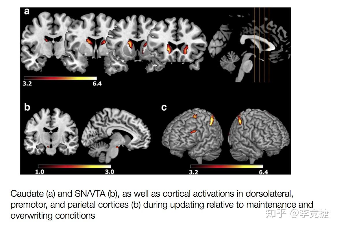
fMRI is a non-invasive imaging technique that measures the metabolic and hemodynamic responses associated with neural activity. It relies on the principle that changes in blood flow to specific regions of the brain correlate with active neural processes. By detecting these changes in blood oxygen level-dependent (BOLD) signals, fMRI provides a high-resolution, real-time view of brain activity.
PET
On the other hand, PET is a nuclear imaging technique that measures the metabolic activity of the brain by tracking the distribution of radiolabeled compounds. PET relies on the injection of a small amount of radioactive glucose, which is metabolized by active brain cells. The emitted gamma rays are detected by PET scanners, providing a detailed map of metabolic activity within the brain.
Comparative Analysis
The primary distinction between fMRI and PET lies in their methodologies for detecting brain activity. fMRI measures changes in blood flow, while PET measures changes in metabolic activity. This difference in approach has implications for the types of information each technique can provide. fMRI is particularly adept at detecting changes in activity associated with sensory processing, motor functions, and cognitive tasks. Its high temporal resolution allows for the observation of rapid changes in brain activity, making it ideal for studying dynamic processes such as language processing and decision-making.
In contrast, PET offers superior spatial resolution and is particularly well-suited for studying the distribution of neurotransmitters and other molecules involved in the regulation of brain function. Its ability to measure metabolic activity directly makes it particularly valuable for studying the effects of drugs and neurological disorders on brain function.
Strengths and Limitations
Both fMRI and PET have their strengths and limitations. fMRI's strengths include its high temporal resolution, non-invasiveness, and ability to detect changes in blood flow associated with neural activity. However, its spatial resolution is generally lower than that of PET, making it less effective for studying small-scale brain structures and processes.

PET, on the other hand, offers superior spatial resolution and the ability to measure metabolic activity directly. However, it is more invasive than fMRI, requiring the injection of radioactive compounds, which can limit its use in certain patient populations.
Applications in Neuroscience and Clinical Practice
The applications of fMRI and PET in neuroscience and clinical practice are vast and varied. fMRI is commonly used in cognitive neuroscience research to study the neural correlates of perception, memory, and language. In clinical practice, fMRI is used to diagnose and monitor brain injuries, neurological disorders, and psychiatric conditions.
PET, on the other hand, is particularly valuable in the study of neurotransmitter function and the effects of drugs on brain activity. It is also used in clinical practice to diagnose and monitor neurological disorders such as Alzheimer's disease, Parkinson's disease, and epilepsy.

In conclusion, fMRI and PET are both indispensable tools in the study of brain function. While fMRI excels in measuring changes in blood flow associated with neural activity, PET offers superior spatial resolution and the ability to measure metabolic activity directly. By combining the strengths of both techniques, neuroscientists and clinicians gain a more comprehensive understanding of the complex processes that underlie brain function. As these technologies continue to evolve, they promise to unlock even greater insights into the mysteries of the human brain.